
Magnification-assisted endodontic care in Indira Nagar
Microscopic Root Canal Treatment in Lucknow
Microscopic root canal treatment uses a dental operating microscope to provide magnification and focused illumination during selected endodontic procedures. It can help the clinician inspect small anatomical details while diagnosing, accessing, cleaning or retreating a root canal system.
At Happi Smiles Dental Clinic and Implant Center in Indira Nagar, Lucknow, Dr Harpreet Singh assesses the tooth first and uses magnification when it can contribute to the planned procedure. A microscope is a clinical tool, not a separate diagnosis or a guarantee of healing.
Microscopic endodontic assessments are available Monday to Sunday, 10:00 AM–9:00 PM. Call to confirm availability.
Medically reviewed by Dr Harpreet Singh on 30 September 2026.
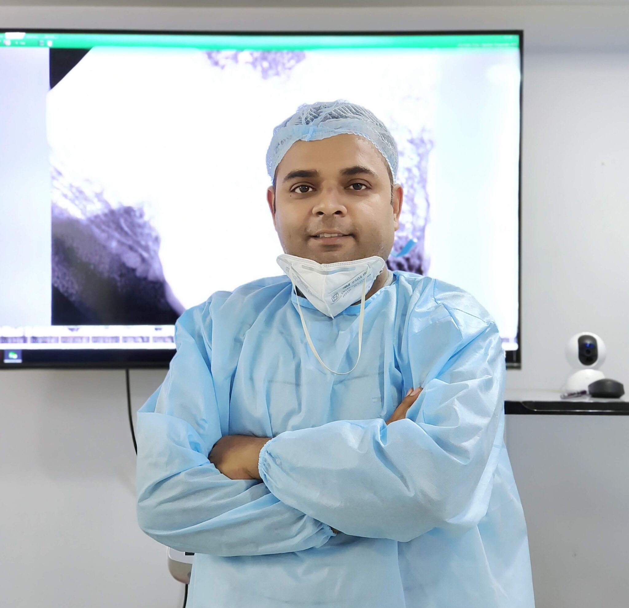
Microscopic Root Canal Treatment in Lucknow: the short answer
Microscopic root canal treatment at Happi Smiles Dental Clinic and Implant Center in Lucknow uses enhanced illumination and magnification during selected endodontic procedures. Dr Harpreet Singh first confirms the diagnosis and tooth outlook because a microscope improves visual access but cannot make every tooth treatable.
A clinical assessment is required before deciding whether this care is appropriate.
What is microscopic root canal treatment?
A dental operating microscope provides coaxial light and adjustable magnification over the treatment field. Endodontic work occurs in small internal spaces, so improved visualisation can assist inspection of the access cavity, pulp chamber floor, canal openings and selected procedural complications.
Microscope assistance enhances visualisation while the success of care still depends on diagnosis, disinfection, sealing and restoration.
Who may need microscopic root canal treatment in Lucknow?
Situations where magnification may help
- Small, hidden or calcified canal openings
- Complex access anatomy
- Assessment during root canal retreatment
- Management of selected obstructions or procedural defects
- Detailed inspection for cracks within the visible field
What the microscope cannot solve
- A non-restorable tooth
- A vertical root fracture with poor outlook
- Severe loss of periodontal support
- Infection in anatomy that remains inaccessible
- A need for surgery or care in another clinical setting
Not every routine tooth requires the same degree of magnification. The clinician selects tools according to anatomy, difficulty and the planned step.
Magnification is most useful when additional visual detail can change how a feasible procedure is performed.
What can a dental operating microscope show in Lucknow?
Focused light and magnification can make the chamber floor, developmental grooves, small canal openings and restorative margins easier to inspect. In retreatment, it may help identify residual material, missed anatomy or the visible end of an obstruction.
Visibility still depends on a direct line of sight. Curved roots and deep canal anatomy extend beyond what can be seen directly, so radiographs, tactile information, electronic measurements and clinical judgment remain necessary. A microscope cannot see through tooth structure.
The microscope improves the visible operative field but must be combined with other diagnostic and endodontic methods.
How is microscopic root canal treatment planned at Happi Smiles?
Dr Harpreet Singh reviews symptoms, medical history, tooth restorability, previous treatment and selected dental images. Pulp and periapical tests help confirm whether root canal treatment is indicated. The clinic reports digital X-rays, intraoral cameras, scanners and advanced training in microscopic endodontics.
Case difficulty includes tooth position, mouth opening, canal curvature, calcification, previous posts or root fillings, cracks and the final restoration. A complex case may still require referral even when magnification is available.
Microscope use follows diagnosis and case-difficulty assessment rather than being applied as a marketing label.
What happens during microscopic root canal treatment?
Positioning
The tooth is numbed, isolated and positioned for stable visual access.
Magnified inspection
The access and visible chamber anatomy are examined under focused light.
Canal treatment
Canals are negotiated, cleaned, shaped and disinfected using the planned protocol.
Seal and restoration
The canal system and access are sealed before definitive restoration.
The microscope can be used for selected parts or throughout the appointment. Treatment time and visits depend on infection, anatomy and whether this is initial care or retreatment.
Magnification supports precise visual steps within a complete endodontic and restorative workflow.
Will I need local anaesthesia in Lucknow?
Microscope-assisted root canal treatment is usually performed with local anaesthesia. The microscope does not touch the tooth or change how anaesthesia works. You may notice that the clinician adjusts your chair and asks you to remain still while the field is viewed.
Tell the clinic about allergies, medicines, pregnancy, heart conditions and previous difficulty becoming numb. Jaw fatigue can occur during longer appointments, so communicate if you need a pause.
Comfort measures are based on the endodontic procedure and patient needs, not on magnification itself.
Aftercare following microscopic root canal treatment
Aftercare is similar to other root canal procedures. The tooth may feel tender for several days, especially if inflammation or infection existed before treatment. Follow the instructions for medicines, hygiene, eating and protection of any temporary restoration.
Contact the clinic for visible swelling, severe pressure lasting more than a few days, an uneven bite, loss of a temporary filling, a suspected medicine reaction or return of earlier symptoms.
Protect the treated tooth and seek review for swelling, worsening pain or loss of its temporary seal.
What should I expect afterwards in Lucknow?
The tooth still requires an appropriate coronal restoration. A filling, onlay or crown may be planned according to the remaining structure and bite. Delaying the final seal can expose the tooth to contamination or fracture.
Healing may be reviewed clinically and with selected radiographs. Enhanced visualisation does not eliminate biological uncertainty, and some teeth later require retreatment, surgery or extraction.
The microscope assists treatment, while final restoration and follow-up remain essential to the tooth’s outlook.
What are the limits of microscopic root canal treatment?
Magnification cannot guarantee that every canal can be negotiated, every separated instrument removed or every crack detected. Some anatomy lies beyond direct vision. Removal of obstructions can also risk weakening or perforating the root.
The clinic may decline or refer care when the tooth cannot be restored, a vertical fracture is suspected, periodontal support is inadequate, surgical treatment is indicated or the case requires equipment or expertise beyond the setting. Alternatives should be discussed without implying that technology alone changes the prognosis.
A microscope improves visual control but does not override anatomy, biology, restorability or procedural risk.
Microscopic Root Canal Treatment at Happi Smiles Dental Clinic in Lucknow
Happi Smiles states that Dr Harpreet Singh has advanced training in microscopic endodontics and that the clinic uses digital imaging and scanning systems. These tools are selected according to the diagnostic question and procedural need.
Magnification with a purpose
The microscope is used when additional visual detail can support the planned step.
Restorability remains central
Technology does not replace assessment of cracks, remaining structure and the final seal.
The clinic combines examination, explanation and access to other dental disciplines when the findings require them.
Meet Dr Harpreet Singh at Happi Smiles
Dr Harpreet Singh completed BDS and MDS training at Manipal in Conservative Dentistry and Endodontics. The clinic reports advanced training in microscopic endodontics. He assesses diagnosis, case difficulty and restorability before deciding how magnification may contribute.
View Dr Harpreet Singh’s profile
Dr Harpreet Singh is identified as the clinician responsible for the page’s general clinical review.
Visiting Happi Smiles Dental Clinic in Indira Nagar
Clinic details
Happi Smiles Dental Clinic and Implant Center
S-95, Bhoothnath Market, Sector 5, Indira Nagar, Lucknow, Uttar Pradesh 226016
Phone: 07019725870
Hours: 10:00 AM–9:00 PM, Monday to Sunday
Hours can change on holidays. Call before travelling or if you need current accessibility or parking guidance.
Clinic map
Load the interactive map only when needed.
The clinic is in Bhoothnath Market, Sector 5, Indira Nagar, Lucknow.
Microscopic Root Canal Treatment FAQs
Is microscopic root canal treatment a different treatment?
The biological treatment goals are the same. The difference is the use of enhanced illumination and magnification during selected diagnostic or procedural steps.
Does every root canal require a microscope?
Not every case has the same visual or technical difficulty. The clinician selects magnification according to anatomy, access and the task being performed.
Can a microscope find every missed canal?
No. It improves inspection of visible anatomy, but canals can be calcified, curved or inaccessible. Imaging and other clinical methods remain necessary.
Can microscopic treatment repair a cracked root?
A microscope may help inspect visible tooth structure, but not every crack can be seen and a vertical root fracture can make the tooth unsuitable for preservation.
Is the procedure more uncomfortable?
The microscope itself does not touch the tooth. Local anaesthesia and isolation are used according to the endodontic procedure, and the chair may be positioned for stable viewing.
Is microscopic treatment useful for retreatment?
It can assist inspection and management of selected altered anatomy, previous materials or obstructions, although retreatment feasibility still depends on procedural risk.
Does magnification assure healing?
No. Healing depends on infection control, anatomy, sealing, restoration, periodontal support and patient factors. No visual aid can assure an outcome.
When should I seek urgent help for endodontic symptoms?
Contact the clinic promptly for swelling, fever, pus or escalating pain. Breathing or swallowing difficulty and swelling extending towards the eye or neck need urgent medical attention.
Individual diagnosis and treatment planning may differ from these general answers.
Arrange an assessment for microscopic root canal treatment in Lucknow
Contact the clinic to discuss symptoms, previous records and current appointment availability.
Contact the clinic
Call or message the clinic. Avoid sending confidential medical records through an unsecured message.
Related dental treatments in Lucknow
Root canal treatment
The core endodontic procedure that magnification may assist.
Explore root canal treatmentRoot canal retreatment
Previously treated teeth may benefit from enhanced inspection during selected retreatment steps.
Explore root canal retreatmentDental crowns
A structurally weakened root-treated tooth may need cuspal protection.
Explore dental crownsClinical information sources
- American Association of Endodontists: Microscopes in endodontics
- AAE position statement: Use of microscopes and magnification devices
- American Association of Endodontists: Root canal treatment
Sources accessed 30 September 2026. This page provides general education and does not replace examination, diagnosis or personalised advice.