Clinically selected dental imaging in Indira Nagar
Dental X-rays in Lucknow
Dental X-rays create images of teeth, roots and surrounding bone that cannot always be evaluated by visual examination alone. The dentist should select the smallest appropriate imaging examination only when the expected diagnostic information can affect care.
At Happi Smiles Dental Clinic and Implant Center in Indira Nagar, Lucknow, Dr Harpreet Singh reviews symptoms, history, examination findings and any usable previous images before recommending dental radiographs.
Diagnostic appointments are available Monday to Sunday, 10:00 AM–9:00 PM. Call to confirm current availability.
Medically reviewed by Dr Harpreet Singh on 30 September 2026.
Dental X-rays in Lucknow: the short answer
Dental X-rays at Happi Smiles Dental Clinic and Implant Center in Lucknow are selected after clinical review to answer a specific diagnostic or treatment-planning question. They are not automatically required at every visit, and previous usable images should be considered before repeating an examination.
A clinical assessment is required before deciding whether this care is appropriate.
What are dental X-rays?
Dental radiographs use a controlled X-ray exposure and an image receptor to show structures that may be hidden during a mouth examination. Depending on the view, an image can help assess tooth decay between teeth, root shape, bone levels, unerupted teeth, infection, trauma or the relationship of nearby anatomical structures.
Two-dimensional views include bitewing, periapical and panoramic images. Cone beam computed tomography creates three-dimensional information but is not a routine substitute for lower-dose two-dimensional imaging. The chosen field of view should match the question being investigated.
The useful dental X-ray is the one selected to answer a defined clinical question with an appropriate field of view.
Who may need dental X-rays in Lucknow?
Possible indications
- Pain, swelling, trauma or unexplained sensitivity
- Suspected decay not fully visible clinically
- Assessment of bone support or tooth roots
- Planning for endodontic, surgical, orthodontic or implant care
- Monitoring a known condition when the result may change management
Reasons to reconsider or defer
- A recent usable image already answers the question
- The requested image would not affect diagnosis or care
- The patient cannot safely cooperate with the selected technique
- A different view or specialist report is required
- The request is based only on a fixed calendar
Age alone does not determine the imaging schedule. Disease risk, dental development, symptoms, prior treatment and examination findings all matter.
Dental imaging should be patient-specific and clinically justified rather than routine.
What problem is being investigated in Lucknow?
Three-dimensional imaging may be considered for selected complex situations, but more information is not automatically better. A broader scan exposes a larger area and may reveal findings requiring interpretation or referral.
Image choice follows the suspected condition and the decision that the result could change.
How are dental X-rays selected at Happi Smiles?
Dr Harpreet Singh reviews the medical and dental history, current symptoms, clinical findings and previous imaging. Happi Smiles reports access to digital dental X-rays and scanning technology. The type and number of views should be limited to those needed for the current assessment.
Tell the clinic if you are or may be pregnant, have difficulty standing or sitting, cannot tolerate an intraoral receptor or have recent images from another dental clinic. Pregnancy does not automatically prevent necessary dental imaging, but it is important information for clinical planning.
Selection combines history, examination, prior records and the minimum imaging needed for the decision.
What happens during dental X-rays?
Positioning
You are positioned for the selected intraoral or extraoral view.
Receptor placement
A small sensor may be placed in the mouth, or the machine may move outside it.
Exposure
You remain still briefly while the image is created.
Review
The dentist checks image quality and interprets it with the clinical findings.
Tell the operator if the receptor causes discomfort or triggers gagging. Repositioning or another suitable technique may help. Retakes should occur only when the first image is not diagnostically usable.
Accurate positioning and a usable first image reduce unnecessary repeat exposure.
How is radiation exposure managed in Lucknow?
Dental imaging uses ionising radiation, so every exposure should be justified and optimised. Current professional recommendations emphasise patient-specific selection, appropriate equipment settings, a field of view limited to the area of interest and avoiding routine or convenience imaging.
Digital technology can support efficient image capture, but “digital” does not remove the need for justification. The benefit of information needed for diagnosis or treatment planning must be weighed against radiation exposure. Ask why an image is recommended and how the result could change care.
Radiation safety begins by avoiding images that are not expected to benefit the patient’s care.
How are dental X-ray findings explained in Lucknow?
The image is interpreted together with symptoms and examination findings. A dark or light area on a radiograph is not a diagnosis by itself. Overlap, angulation, restorations and normal anatomy can affect appearance, and some early disease may not be visible.
The dentist should explain the relevant finding, its level of certainty and whether monitoring, another view, treatment or referral is advised. Complex scans or unexpected findings may require review by an oral and maxillofacial radiologist.
Radiographic findings support clinical judgment; they do not replace history and examination.
What should I expect afterwards in Lucknow?
Routine dental imaging does not usually require recovery time. The important next step is understanding what the image did or did not show. Ask whether a copy can be shared with another treating dentist when continuity of care requires it.
Keep details of recent images so another clinic can consider them before ordering repeats. A new image may still be necessary when symptoms, anatomy, image quality or the clinical question differs from the earlier examination.
Good record sharing can reduce avoidable repetition while preserving necessary diagnostic imaging.
What are the limits of dental X-rays?
Radiographs cannot show every crack, early cavity, soft-tissue condition or source of pain. Two-dimensional images compress three-dimensional anatomy and may hide structures through overlap. An image also cannot determine symptoms, tenderness or tooth vitality without clinical tests.
The clinic may decline an image requested without clinical justification, postpone imaging if safe positioning is not possible or recommend a different facility when specialist equipment or interpretation is required. Refusing a necessary diagnostic image can also limit the dentist’s ability to diagnose or treat safely.
Imaging has defined strengths and limitations, so the test must fit the question and the clinical setting.
Dental X-rays at Happi Smiles Dental Clinic in Lucknow
Happi Smiles reports digital X-rays, intraoral cameras and scanning systems. These tools serve different purposes: a camera records visible surfaces, while radiographs can show selected hard-tissue structures that examination cannot directly reveal.
Clinical justification
Images are linked to a diagnostic or planning question.
Review in context
Radiographic findings are interpreted alongside history and examination.
The clinic combines examination, explanation and access to other dental disciplines when the findings require them.
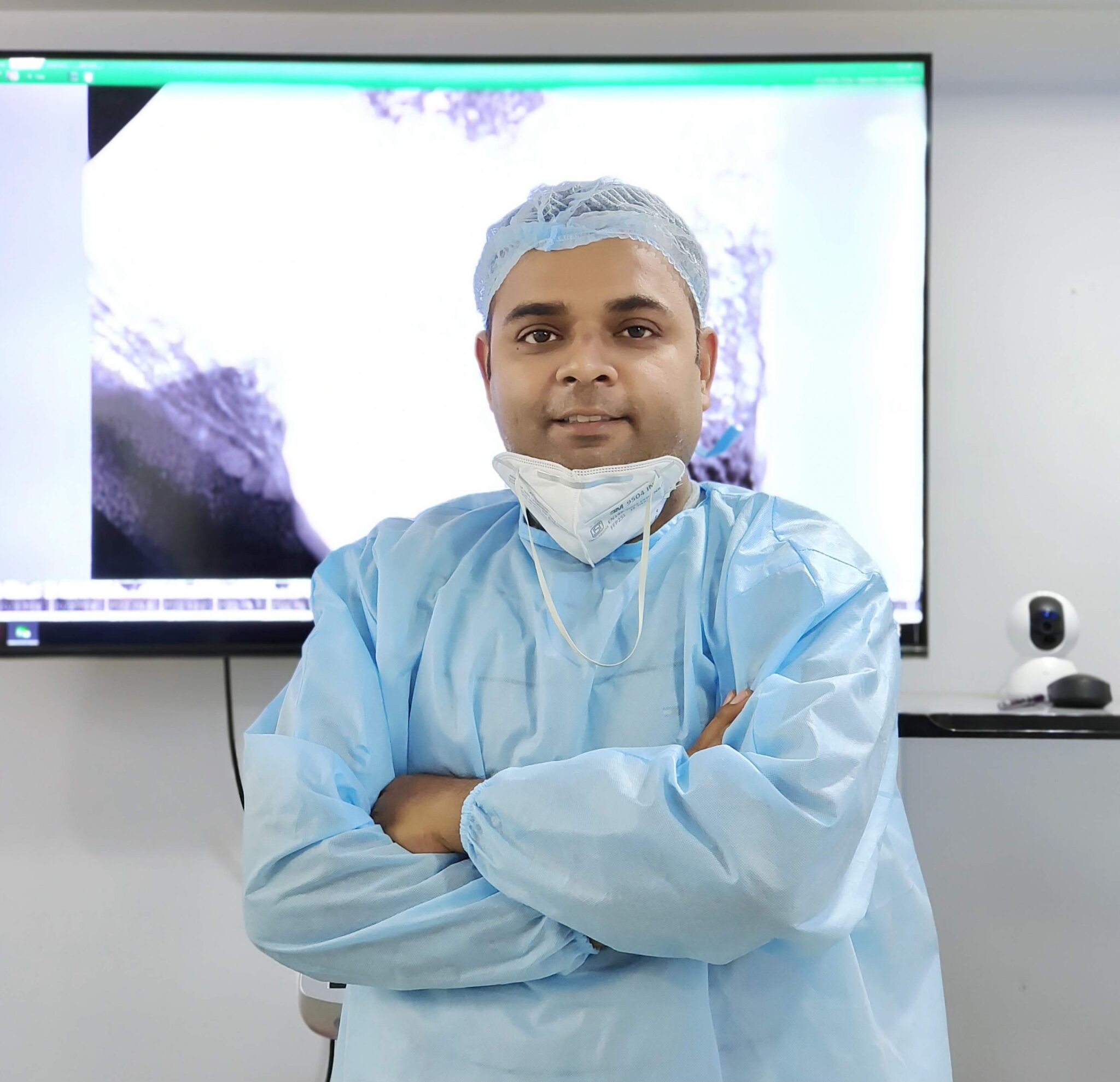
Meet Dr Harpreet Singh at Happi Smiles
Dr Harpreet Singh completed BDS and MDS training at Manipal in Conservative Dentistry and Endodontics, with training in microscopic endodontics reported by the clinic. He uses dental imaging when it contributes to diagnosis, treatment planning or review and discusses the finding in clinical context.
View Dr Harpreet Singh’s profile
Dr Harpreet Singh is identified as the clinician responsible for the page’s general clinical review.
Visiting Happi Smiles Dental Clinic in Indira Nagar
Clinic details
Happi Smiles Dental Clinic and Implant Center
S-95, Bhoothnath Market, Sector 5, Indira Nagar, Lucknow, Uttar Pradesh 226016
Phone: 07019725870
Hours: 10:00 AM–9:00 PM, Monday to Sunday
Hours can change on holidays. Call before travelling or if you need current accessibility or parking guidance.
Clinic map
Load the interactive map only when needed.
The clinic is in Bhoothnath Market, Sector 5, Indira Nagar, Lucknow.
Dental X-rays FAQs
Are dental X-rays needed at every check-up?
No. Current recommendations support patient-specific selection using history, previous images, disease risk, symptoms and clinical findings rather than automatic imaging at every visit.
What is the difference between bitewing and periapical X-rays?
Bitewings commonly show the crowns of upper and lower back teeth and nearby bone. Periapical views show a selected tooth from crown to root tip and surrounding bone.
Is a panoramic X-ray the same as CBCT?
No. A panoramic radiograph is a two-dimensional overview. CBCT creates three-dimensional information and should be reserved for situations where that additional information is justified.
Should I tell the dentist if I may be pregnant?
Yes. Share confirmed or possible pregnancy before imaging so the dentist can consider urgency, benefits, technique and current professional recommendations for your situation.
Can I bring dental X-rays from another clinic?
Yes. Recent diagnostic-quality images may be useful. The dentist will decide whether they answer the current question or whether another view is clinically necessary.
Why might an X-ray need to be repeated?
A repeat may be required if movement, positioning, overlap or another technical problem makes the first image unsuitable for diagnosis. Repeats should not be routine.
Can dental X-rays find every cause of tooth pain?
No. Some cracks, pulp conditions, bite problems and soft-tissue causes require examination and other tests. A normal-looking radiograph does not automatically rule out disease.
When is urgent care needed instead of waiting for imaging?
Seek prompt assessment for severe pain, swelling, fever, trauma or uncontrolled bleeding. Difficulty breathing or swallowing and rapidly spreading swelling need urgent medical attention.
Individual diagnosis and treatment planning may differ from these general answers.
Arrange an assessment for dental x-rays in Lucknow
Contact the clinic to discuss symptoms, previous records and current appointment availability.
Contact the clinic
Call or message the clinic. Avoid sending confidential medical records through an unsecured message.
Related dental treatments in Lucknow
Dental check-up
Clinical examination establishes whether diagnostic imaging is indicated.
Explore dental check-upDental fillings
Selected X-rays can support assessment of decay before restorative care.
Explore dental fillingsRoot canal treatment
Periapical imaging can contribute to endodontic diagnosis and treatment review.
Explore root canal treatmentClinical information sources
- ADA: 2026 dental X-ray recommendations
- ADA: Radiographic imaging
- ADA MouthHealthy: Dental radiographs, benefits and safety
Sources accessed 30 September 2026. This page provides general education and does not replace examination, diagnosis or personalised advice.